Babyecho Fundamentals Explained
Wiki Article
An Unbiased View of Babyecho
Table of ContentsSome Ideas on Babyecho You Need To KnowLittle Known Questions About Babyecho.Indicators on Babyecho You Need To KnowThe 9-Minute Rule for BabyechoSee This Report about BabyechoExcitement About BabyechoWhat Does Babyecho Mean?Babyecho Things To Know Before You Get ThisA Biased View of BabyechoHow Babyecho can Save You Time, Stress, and Money.9 Simple Techniques For Babyecho
It's supplied to everybody and is covered by many insurance coverage strategies. This genetic screening ultrasound is optional. Throughout this ultrasound, your physician will certainly try to find signs of chromosomal disorders. Chromosomal conditions mean that the baby obtained an additional chromosome at conception and might have moderate to severe physical or mental difficulties.
Not known Details About Babyecho
Would I end my pregnancy if there was a danger of Downs Disorder, Trisomy 13, Trisomy 18, or other hereditary disorder? Would recognizing about my pregnancy's danger of hereditary problem make it easier to mentally or literally prepare for a baby with a birth problem? Would certainly it be easier for me to deal with and appreciate pregnancy if I concentrate on the more most likely positive result rather than the opportunity of birth defects?If you're still uncertain, you can review the advantages and disadvantages with your OBGYN. This is the ultrasound that individuals anticipate one of the most! The full makeup ultrasound is usually executed at about 20 weeks, or 5 months. As the name implies, this ultrasound will check out all the child's organ systems to make sure they exist, are a regular dimension and form, and remain in the right area.
Excitement About Babyecho
An extra-small infant or an infant who does not expand according to their development curve could mean that the child is not getting enough nutrition through the placenta and might require to be provided early. 2D ultrasounds are the black and white images that you're probably made use of to seeing. To an inexperienced eye, they can look quite fuzzy or obscure.The best area to have an ultrasound done is always at a clinic, where you will certainly have accessibility to a doctor that has been educated in translating the pictures. At Madison Female's Health, we more than happy to publish off pictures for you to place in your child's scrapbook or anywhere else you want to present those "coming soon" images.
How Babyecho can Save You Time, Stress, and Money.
:max_bytes(150000):strip_icc()/191127-ultrasound-trimester-pink-2000-fd089add04f8444e9d7a403933d1994f.jpg)
A handheld probe will certainly after that be conformed the area. The gel helps the probe send acoustic waves. These waves bounce off the body structures, consisting of the establishing baby, to create an image on the ultrasound machine. Sometimes, a pregnancy ultrasound might be done by positioning the probe into the vaginal canal.
Babyecho - An Overview
You will certainly need to have a full bladder to obtain the finest ultrasound photo. You may be asked to drink 2 to 3 glasses of liquid an hour prior to the examination. https://hubpages.com/@babydoppler1.Some centers are currently executing a maternity ultrasound called a nuchal clarity testing examination around 9 to 13 weeks of maternity. This test is done to look for signs of Down disorder or other issues in the creating infant.
Little Known Questions About Babyecho.
After a favorable pregnancy examination outcome, you will need to validate your pregnancy is viable by ultrasound. A viable maternity is one that is expected to continue and cause childbirth (if nothing else actions are taken). If no fetal heart beat is discovered, you would not require an abortion but would certainly be referred for medical treatment rather.As a freshly expectant mom, you're possibly excited to have your child's first ultrasound. Listed below, we've damaged down what an ultrasound is and what your medical professional will be looking for throughout the maternity.
Audio waves go through your uterus, then bounce off the infant as resonances. This makes ultrasound very preferable for pregnancy.
The Facts About Babyecho Uncovered
It is a wand-shaped probe, generally covered with a condom and lube, then inserted into the vaginal area. The ultrasound technician will relocate the tool to understand they need on the screen. This sort of ultrasound is not taken into consideration painful but might be uncomfortable. The technician might utilize this technique if you need an ultrasound early in the maternity, while the unborn child is deep within the mom's pelvis.The transvaginal probe may additionally be utilized later in maternity to measure the placenta or size of the cervix. The stomach ultrasound is what many individuals think about when it concerns ultrasounds for infants. The service technician uses clear gel to your stomach, then moves the transductor along your abdominal area.
An Unbiased View of Babyecho
You might not hear the infant's heartbeat at the initial trimester ultrasound visit, which's alright. It might be because you're not far enough along in the maternity or the baby's setting. Sometimes the infant's heart beat can be discovered as early as 5 to 6 weeks after perception. You can normally hear the heartbeat much better closer to 10 weeks after pregnancy.Your physician can inspect the infant's size utilizing ultrasound innovation. In the initial trimester, they can examine the crown-to-rump size. This primarily aids them determine how far along the baby is.
The Single Strategy To Use For Babyecho
Your doctor may advise that you have 1 or more ultrasounds at different factors in your maternity. Depending on how much along your pregnancy is, ultrasound photos aid your doctor: estimate your due day check points like the dimension and position of the fetus to make certain everything is normal see the position of the placenta see the quantity of amniotic fluid in your uterus discover numerous pregnancies (twins, triplets, and article source so on) Ultrasounds can additionally be utilized to evaluate for particular birth problems, like Down disorder.
It feels like a routine genital test that you might get throughout a wellness visit. You could really feel a bit of pressure, yet it's not uncomfortable. Doctors, midwives, or educated ultrasound specialists will do your ultrasound and read the outcomes. The expense of an ultrasound relies on the sort of ultrasound you obtain and where you get it.
The Single Strategy To Use For Babyecho

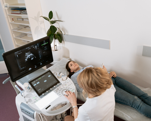
The darkness makes it easier for the person doing the check to plainly see the picture on the screen. You might require to roll down the waistband of your trousers.
Not known Facts About Babyecho
This is to protect your clothing from the ultrasound gel. Next, they will put the ultrasound gel onto your stomach. The gel assists the probe to relocate. As the probe conforms your belly, a photo will certainly show up on the display. The pictures you see are in black and white.You might not be able to inform where your child is in the picture. The person doing the scan will frequently point things out to you like the baby's heartbeat and head.
Report this wiki page